U.S. flag

An official website of the United States government

NCBI Bookshelf. A service of the National Library of Medicine, National Institutes of Health.

Adam MP, Feldman J, Mirzaa GM, et al., editors. GeneReviews® [Internet]. Seattle (WA): University of Washington, Seattle; 1993-2024.

  • This publication is provided for historical reference only and the information may be out of date.

This publication is provided for historical reference only and the information may be out of date.

Cover of GeneReviews®

GeneReviews® [Internet].

Show details

Central Core Disease – RETIRED CHAPTER, FOR HISTORICAL REFERENCE ONLY

, MD, PhD and , MD, PhD.

Author Information and Affiliations

Initial Posting: ; Last Update: December 4, 2014.

Estimated reading time: 32 minutes

Summary

NOTE: THIS PUBLICATION HAS BEEN RETIRED. THIS ARCHIVAL VERSION IS FOR HISTORICAL REFERENCE ONLY, AND THE INFORMATION MAY BE OUT OF DATE.

Clinical characteristics.

Central core disease (CCD) is characterized by muscle weakness ranging from mild to severe. Most affected individuals have mild disease with symmetric proximal muscle weakness and variable involvement of facial and neck muscles. The extraocular muscles are often spared. Motor development is usually delayed, but in general, most affected individuals acquire independent ambulation. Life span is usually normal. Severe disease is early in onset with profound hypotonia often accompanied by poor fetal movement, spinal deformities, hip dislocation, joint contractures, poor suck, and respiratory insufficiency requiring assisted ventilation. The outcome ranges from death in infancy to survival beyond age five years. The weakness in CCD is not typically progressive.

Diagnosis/testing.

The diagnosis of CCD is based on clinical findings of muscle weakness, the histopathologic findings of characteristic cores on muscle biopsy, and molecular genetic testing. Most CCD is associated with pathogenic variants in RYR1, the gene encoding the ryanodine receptor 1.

Management.

Treatment of manifestations: Physical therapy for hypotonia and weakness that may include stretching and mild to moderate low-impact exercise; assistive devices as needed for ambulation; orthopedic surgery as needed for scoliosis, congenital hip dislocation, foot deformities; respiratory support, breathing exercises, chest physiotherapy as needed; dietary supplementation and nasogastric or gastrostomy feeding as needed.

Prevention of secondary complications: Intervention as needed to prevent respiratory compromise from scoliosis; immunization against influenza; prompt treatment of respiratory infection; mobility and physical therapy to prevent joint contractures.

Surveillance: Routine assessment of spine for scoliosis, joints for contractures, respiratory parameters (e.g., respiratory rate, peak expiratory flow rate [PEFR], forced vital capacity [FVC], and forced expiratory volume in one second [FEV1]), motor abilities to determine need for physical therapy, occupational therapy, assistive devices; sleep studies when signs of nocturnal hypoxia are present.

Agents/circumstances to avoid: Although the actual risk for malignant hyperthermia susceptibility is unknown, it is prudent for individuals with CCD to avoid inhalational anesthetics and succinylcholine.

Evaluation of relatives at risk: If the RYR1 pathogenic variant is known, it is appropriate to offer at-risk relatives molecular genetic testing to identify those with possible increased malignant hyperthermia susceptibility.

Genetic counseling.

Central core disease (CCD) is usually inherited in an autosomal dominant (AD) manner but can be inherited in an autosomal recessive (AR) manner. Most individuals diagnosed with AD central core disease have an affected parent or an asymptomatic parent who has a pathogenic variant. The proportion of AD CCD caused by de novo pathogenic variants is unknown. Each child of an individual with AD CCD has a 50% chance of inheriting the pathogenic variant. The parents of a child with AR CCD are obligate heterozygotes and therefore carry one mutated allele. Heterozygotes (carriers) are often asymptomatic. At conception, each sib of an individual with AR CCD has a 25% chance of being affected, a 50% chance of being an asymptomatic carrier, and a 25% chance of being unaffected and not a carrier. Prenatal diagnosis for pregnancies at increased risk for AD or AR CCD is possible once the pathogenic variant(s) have been identified in an affected family member.

Diagnosis

Suggestive Findings

Diagnosis of central core disease should be suspected in individuals with the following features – keeping in mind that this disorder has a wide spectrum of symptoms and presentations, from the absence of symptoms to severe features necessitating the use of ventilatory support:

  • In early-onset disease:
    • Hypotonia and generalized weakness, often accompanied by perinatal complications including poor fetal movement, respiratory insufficiency, and poor suck
    • Delayed motor milestones (Independent ambulation is commonly achieved between ages three and four years, but varies depending on the severity of the disease.)
    • Spinal deformities, congenital hip dislocation, high-arched palate, foot deformities, and joint contractures. Rarely, patients may show severe skeletal malformations such as those seen in spondylocostal dysostosis.
  • In later-onset disease (rare):
    • Mild symmetric myopathy, predominantly involving the proximal muscles
    • Mildly affected facial muscles
    • Occasional involvement of the extraocular muscles (Ophthalmoplegia is relatively common in the autosomal recessive forms.)

Establishing the Diagnosis

The diagnosis of central core disease is established in a proband with (1) a muscle biopsy demonstrating characteristic cores in a significant number of fibers on muscle biopsy, even in individuals who are seemingly asymptomatic and/or (2) molecular genetic testing that detects pathogenic variant(s) in RYR1 (see Table 1).

In the event that molecular testing is done first:

  • In a patient with classic clinical presentation of CCD, if the identified RYR1 variant is known to be pathogenic, the necessity of muscle biopsy becomes less important and will only be used for classification.
  • In a patient without visible clinical involvement, muscle biopsy will be of importance if there is a need to interpret RYR1 variations of uncertain significance, in the absence of functional studies.

Muscle biopsy

Histologic examination of muscle is essential to the diagnosis of central core disease. Diagnostic findings are the presence of a significant number of cores in type 1 fibers with the following characteristics (Figure 1B):

Figure 1.

Figure 1.

Histologic features of muscle observed in central core disease A-B. Sections from a child age nine years depicting the classic description of CCD

  • Often well demarcated
  • May be centrally or peripherally located in the fibers
  • Run down an appreciable length of the fiber on longitudinal sections
  • Devoid of mitochondria
  • Do not stain with oxidative enzyme stains (e.g., NADH-tetrazolium reductase, succinate dehydrogenase, cytochrome c oxidase)
  • Deficient in phosphorylase activity and glycogen
  • Sometimes surrounded by a thin rim of high oxidative enzyme activity, giving the appearance of "rimmed cores"
  • On immunohistochemistry studies, distinct staining patterns that are restricted to the cores: RyR1 protein was focally depleted within the cores, while other proteins including DHPR[alpha]1s, triadin, SERCA1/2, and calsequestrin accumulated within or around the cores [Herasse et al 2007].

Less common but nonetheless important pathologic findings in the spectrum of cores include the following [Ferreiro et al 2002b, Jungbluth et al 2002, Sewry et al 2002]:

  • More than one core can be observed within a single muscle fiber.
  • The number of type 1 fibers with cores varies.
  • The diameter of cores can vary.
  • Foci of multiple minicores in focal areas can occur.

Other pathologic characteristics of muscle:

  • Type 1 fiber predominance or uniformity
  • Mild to moderate fiber size variation
  • Minimal to moderate endomysial fibrosis. Marked fibrosis and increase in adipose tissue have been noted in several cases.
  • Occasional increase in internal and central nuclei

Note: (1) Nemaline bodies occurring together with cores have been seen in genetically confirmed cases of CCD. When rods are numerous this has sometimes been referred to as core-rod disease. In a large French pedigree demonstrating autosomal dominant inheritance, the association of this disease with RYR1 pathogenic variants was confirmed [Monnier et al 2000]. Interestingly, some cases of nemaline myopathy may also show cores [Jungbluth et al 2002], blurring the pathologic distinction between the two disorders. (2) Facial muscle involvement and high-arched palate are almost always observed in infantile or childhood nemaline myopathy, but are rarely seen in CCD.

Ultrastructural studies show the following:

  • Virtual absence of mitochondria and sarcoplasmic reticulum (SR) in the core region. SR accumulation within the cores has been described on EM.
  • Irregular zigzag pattern or complete disruption of the Z-lines but often preservation of the striation pattern
  • Reduction in the intermyofibrillar space

Molecular Genetic Testing

One genetic testing strategy is single-gene testing. Sequence analysis of RYR1 is performed first, and followed by deletion/duplication analysis if no pathogenic variant is found (see Table 1).

An alternative genetic testing strategy is use of a multigene panel that includes RYR1 and other genes of interest (see Differential Diagnosis). Note: (1) The genes included in the panel and the diagnostic sensitivity of the testing used for each gene vary by laboratory and are likely to change over time. (2) Some multigene panels may include genes not associated with the condition discussed in this GeneReview; thus, clinicians need to determine which multigene panel is most likely to identify the genetic cause of the condition at the most reasonable cost while limiting identification of variants of uncertain significance and pathogenic variants in genes that do not explain the underlying phenotype. (3) In some laboratories, panel options may include a custom laboratory-designed panel and/or custom phenotype-focused exome analysis that includes genes specified by the clinician. (4) Methods used in a panel may include sequence analysis, deletion/duplication analysis, and/or other non-sequencing-based tests.

For an introduction to multigene panels click here. More detailed information for clinicians ordering genetic tests can be found here.

Note: Although in the majority of patients CCD results from a single pathogenic variant in RYR1, individuals with biallelic pathogenic variants (autosomal recessive inheritance) have been reported; thus, the following considerations are important.

If only one pathogenic variant is identified in a simplex case (i.e., a single occurrence in a family), it is difficult to distinguish between the following:

To resolve this issue, the following can be considered:

More comprehensive genomic testing. If serial single-gene testing (and/or use of a multigene panel) has not confirmed a diagnosis in an individual with features of CCD, genomic testing may be considered. Such testing may include exome sequencing, genome sequencing, and mitochondrial sequencing.

For an introduction to comprehensive genomic testing click here. More detailed information for clinicians ordering genomic testing can be found here.

Table 1.

Molecular Genetic Testing Used in Central Core Disease

Gene 1MethodProportion of Probands with a Pathogenic Variant Detectable by Method
RYR1 Sequence analysis of select exons 2, 347%-80% 4
Sequence analysis of entire RYR1 2>90% 5
Deletion/duplication analysis 6Unknown
Unknown 7NA
1.

See Table A. Genes and Databases for chromosome locus and protein. See Molecular Genetics for information on allelic variants detected in this gene.

2.

Sequence analysis detects variants that are benign, likely benign, of uncertain significance, likely pathogenic, or pathogenic. Pathogenic variants may include small intragenic deletions/insertions and missense, nonsense, and splice site variants; typically, exon or whole-gene deletions/duplications are not detected. For issues to consider in interpretation of sequence analysis results, click here.

3.

Exons sequenced vary by laboratory.

4.
5.

Results from Wu et al [2006]. Among 27 individuals diagnosed with CCD on muscle biopsy, sequence analysis of the entire coding region documented RYR1 pathogenic variants in 93% [Wu et al 2006], suggesting that CCD may not be a genetically heterogeneous disease, as previously thought.

6.

Testing that identifies exon or whole-gene deletions/duplications not detectable by sequence analysis of the coding and flanking intronic regions of genomic DNA. Methods used may include quantitative PCR, long-range PCR, multiplex ligation-dependent probe amplification (MLPA), and chromosomal microarray (CMA) that includes this gene/chromosome segment.

7.

No other locus heterogeneity has been identified. It is possible that other disorders with EC uncoupling could be within the spectrum of CCD, but more studies are warranted. Other candidate genes to be considered include those that code for proteins involved or associated with the triadin, which is the anatomic site of EC uncoupling, and include triadin, junctin, histidine-rich calcium-binding protein, calsequestrin, JP-45, and mitsugamin-29 [Treves et al 2005] and dihydropyridine receptor, calmodulin, and inositol phospate 3 receptor. To date, no pathogenic variant in the genes encoding these proteins has been associated with CCD.

Clinical Characteristics

Clinical Description

The expressivity of central core disease (CCD) is variable even among family members, ranging clinically from mild (i.e., almost asymptomatic) to severe (i.e., ventilator-dependent) and histologically varying in the extent and localization of cores in the muscle fibers.

Mild disease. Most individuals have mild disease characterized by mild, symmetric weakness that preferentially affects the proximal muscles. The facial and neck muscles may be mildly involved in some cases. The extraocular muscles are often spared in the classic, autosomal dominant form, but are typically involved in the autosomal recessive form. Motor development is usually delayed, but in general, most affected individuals acquire independent ambulation. Hypotonia in infancy and respiratory insufficiency can also occur in those with mild disease. Life span is usually normal.

Muscle cramps have been documented in some individuals with CCD, and this may be associated with MH susceptibility.

Severe disease is characterized by infantile onset associated with profound hypotonia and respiratory dysfunction requiring continuous assisted ventilation. In severely affected individuals, death may result from respiratory infection or respiratory insufficiency.

Fetal akinesia has been associated with both autosomal dominant and autosomal recessive forms of RYR1-related CCD [Romero et al 2003]. The clinical phenotype consisted of severe hypotonia, arthrogryposis multiplex congenita, amyotrophy, and respiratory failure, requiring mechanical ventilation. The outcome, however, was variable (ranging from early death to survival beyond age five years).

Progression. CCD is not typically progressive, although slow progression has been reported. Scoliosis can be progressive, resulting in respiratory insufficiency.

Cognition. Intellectual ability is intact.

Laboratory findings

  • Serum creatine kinase concentration may be normal or mildly elevated.
  • Electromyography may confirm the presence of myopathy and reveal brief, short action potentials and early recruitment.

Muscle magnetic resonance imaging. Muscle imaging has demonstrated that certain muscles are selectively and consistently involved in autosomal dominant RYR1-related myopathies, including quadriceps, sartorius, adductor magnus, soleus, gastrocnemius, and peroneal group; certain muscles are relatively spared, including rectus femoris, gracilis, adductor longus, and tibialis anterior [Jungbluth et al 2004]. These findings were supported by Fischer et al [2006] who described distinct MRI findings in persons with CCD who have an RYR1 pathogenic variant, including predominant involvement of the gluteus maximus, adductor magnus, sartorius, vastus intermediolateralis, soleus, and lateral gastrocnemius muscles, as compared to those who do not have an RYR1 pathogenic variant. The rectus femoris, adductor longus, and gracilis are hypertrophied in some patients. MRI findings in autosomal recessive RYR1-related CCD are not as consistent.

Genotype-Phenotype Correlations

Although most RYR1 pathogenic variants that result in CCD are inherited in an autosomal dominant manner, reports of autosomal recessive inheritance are increasing. It is not possible to predict the mode of inheritance based on the pathogenic variant alone.

Some studies have shown that autosomal recessive CCD, often associated with RYR1 pathogenic variants outside the C-terminal region, can be severe [Romero et al 2003, Zhou et al 2006b]. Thus, it may be possible to consider most autosomal dominant forms of CCD as milder in phenotype than autosomal recessive forms of CCD.

In a study of 25 individuals with genetically confirmed CCD, Wu et al [2006] determined that:

  • The 16 individuals with C-terminal RYR1 pathogenic variants had certain clinical features including hypotonia during infancy, delayed motor development, and limb muscle weakness and certain pathologic findings on muscle biopsy that delineate C terminal pathogenic variants from other groups including (1) type 2 fiber deficiency and interstitial fibrosis, (2) characteristic cores with clearly demarcated borders that are observed in almost all type 1 muscle fibers, (3) higher than average frequency of "rimming" on the borders of these cores;
  • Most individuals with CCD with at least one RYR1 pathogenic variant outside the C-terminal region had only mild musculoskeletal abnormalities such as joint contractures and scoliosis. Inheritance was autosomal dominant, consistent with previous reports of mild CCD phenotype.

Malignant hyperthermia susceptibility (MHS)-related RYR1 pathogenic variants are predominantly located in the hydrophilic N-terminal and central portions of the ryanodine receptor 1 (RyR1) protein, whereas CCD-related RYR1 substitutions mainly occur in the hydrophobic pore-forming region of the channel [Monnier et al 2000, Monnier et al 2001, Davis et al 2003, Zorzato et al 2003]. Previous reports have asserted that persons without muscle disease who are susceptible to malignant hyperthermia (MH) have pathogenic variants in the C-terminal region of ryanodine receptor 1; however, limited histopathologic evaluation of these individuals has revealed the presence of cores that are not characteristic of the cores of CCD [Ibarra et al 2006]; thus, they are most appropriately labeled as having "MH with cores."

Individuals with CCD who have pathogenic variants in the N-terminal domain may have a higher probability of malignant hyperthermia susceptibility than those with pathogenic variants in the C-terminal domain [Wu et al 2006].

Penetrance

In general, the penetrance of CCD-associated RYR1 pathogenic variants is almost 100%. Clinical variability exists to some extent among affected members of the family.

Nomenclature

CCD has also been referred to as Shy-Magee syndrome, after the individuals who initially reported it.

Some cases called core-rod disease are not associated with a RYR1 pathogenic variant; thus, "core-rod disease" is not a true synonym for CCD.

Prevalence

An accurate incidence and prevalence of CCD, considered to be the most frequently occurring congenital myopathy, are unknown. By definition, CCD is considered a rare disease by the Office of Rare Disease, implying that it affects fewer than 200,000 in the US population. A recent estimate of 1:100,000 was calculated among patients with genetic muscle disease in Northern England [Norwood et al 2009]. In Japan, the carrier frequency for heterozygous RYR1 pathogenic variants is reported to be as high as 1:2,000 [Wu et al 2006].

Differential Diagnosis

The clinical findings of central core disease (CCD) are variable and not disease specific; they can be seen in other congenital myopathies. Thus, from a clinical standpoint CCD cannot be readily distinguished from other congenital myopathies, such as multiminicore disease (OMIM 606210, 180901), CNMDU1 (see Allelic Disorders), the intermediate form of nemaline myopathy, fingerprint body myopathy (OMIM 305550), congenital fiber-type disproportion (OMIM 255310), hyaline body myopathy (OMIM 255160), reducing body myopathy (OMIM 300717, 300718), and cylindrical spirals myopathy (OMIM 160990).

The "central core" histologic changes are nonspecific and may occur in other myopathies. Cores that have been noted in CCD have also been reported with pathogenic variants in the following genes:

  • SELENON (SEPN1). Pathogenic variants in this gene are also associated with minicores [Ferreiro et al 2002b], but no individual with a SELENON pathogenic variant and the typical long, well-delimited central cores characteristic of CCD has been reported.
  • MYH7, in hypertrophic cardiomyopathy
  • ACTA1 and TNNT1 in nemaline myopathy [Ilkovski et al 2001]. ACTA1 pathogenic variants were found in a congenital myopathy with few cores on muscle biopsy [Kaindl et al 2004]; like other disorders with cores, however, these disorders are better considered as myopathies with cores, not CCD.
  • CFL2, encoding cofilin-2, has been associated with nemaline myopathy with minicores [Agrawal et al 2007].
  • Structures similar to cores have been observed in the myofibers of individuals with neurogenic atrophy but are more appropriately called "target fibers" in this setting because of the darker band around the pale central area, giving it a target-like appearance. In addition, core-like lesions devoid of this band can also be seen conditions with neurogenic atrophy.

Management

Evaluations Following Initial Diagnosis

To establish the extent of disease and needs in an individual diagnosed with central core disease (CCD), the following evaluations are recommended:

  • Neurologic examination with attention to features of congenital myopathy (hypotonia, failure to thrive, joint contractures, scoliosis), weakness of the limbs, and muscle cramps
  • Physical and occupational therapy assessments
  • Evaluation for feeding difficulties, including assessment for sucking and ability to swallow
  • Pulmonary function testing in most patients, especially those with scoliosis, hypotonia, signs of respiratory distress, and/or history of recurrent chest infections. History should be taken for symptoms of nocturnal hypoxia including early morning headaches, daytime drowsiness, loss of appetite, and deteriorating school performance.
  • Consultation with a clinical geneticist and/or genetic counselor

Treatment of Manifestations

Since prognosis is mainly influenced by respiratory status and scoliosis, treatment geared toward these manifestations is essential.

Treatment depends on the severity of symptoms, but mainly consists of supportive measures and rehabilitation that address the following problems:

  • Hypotonia and weakness. Patients may benefit from physical therapy. Interventions may include stretching programs and mild to moderate low-impact exercise; activities should be balanced in such a way that exhaustion is avoided.
  • Scoliosis and joint contractures. Some patients may only require physical therapy, while others may need orthopedic surgery (e.g., scoliosis surgery, corrective surgery for congenital hip dislocation and foot deformities).
  • Respiratory. Patients with more severe symptoms may require respiratory support. Breathing exercises and chest physiotherapy for handling secretions may also be beneficial.
  • Feeding difficulties. Individuals may require diet supplementation and feeding by means of nasogastric/orogastric routes or gastrostomy.

Prevention of Secondary Complications

Secondary complications can include respiratory compromise from scoliosis; hence, orthopedic intervention may reduce the risk of this problem.

Immunization against influenza is encouraged.

Prompt treatment of respiratory infection is important.

Joint contractures may be prevented by encouraging mobility and by active participation in physical therapy.

Surveillance

The following are appropriate:

  • Routine assessment of the spine for scoliosis and joints for contractures
  • Routine assessment of respiratory parameters such as respiratory rate, peak expiratory flow rate (PEFR), forced vital capacity (FVC), and forced expiratory volume in one second (FEV1)
  • Sleep studies especially when patients show signs of nocturnal hypoxia
  • Regular assessment of motor abilities in order to determine need for physical therapy, occupational therapy, and assistive devices for ambulation, such as a wheelchair

Agents/Circumstances to Avoid

Although it is unknown how CCD is associated with malignant hyperthermia susceptibility or which pathogenic variants in RYR1 are absolutely related to MH susceptibility, it is prudent for individuals with CCD to avoid inhalational anesthetics and succinylcholine. See Malignant Hyperthermia Susceptibility for more details.

Individuals suspected of having MH susceptibility are advised to avoid extremes of heat, but this does not mean restriction of athletic activity.

Evaluation of Relatives at Risk

Because CCD is associated with an increased risk for MH susceptibility, it is appropriate to test at-risk relatives of a proband (whether symptomatic or not) for the RYR1 pathogenic variant identified in the proband in order to caution those with the pathogenic variant about potential risks of inhalational anesthetics and succinylcholine. See Malignant Hyperthermia Susceptibility for more details.

See Genetic Counseling for issues related to testing of at-risk relatives for genetic counseling purposes.

Pregnancy Management

Because all patients with CCD are considered to be at risk for malignant hyperthermia, inhalational anesthetics and succinylcholine should be avoided while considering general or regional anesthesia for delivery. Similar to most neuromuscular disorders, clinical progression of disease can be expected in CCD [Sax & Rosenbaum 2006].

Therapies Under Investigation

Search ClinicalTrials.gov in the US and EU Clinical Trials Register in Europe for access to information on clinical studies for a wide range of diseases and conditions. Note: There may not be clinical trials for this disorder.

Genetic Counseling

Genetic counseling is the process of providing individuals and families with information on the nature, mode(s) of inheritance, and implications of genetic disorders to help them make informed medical and personal decisions. The following section deals with genetic risk assessment and the use of family history and genetic testing to clarify genetic status for family members; it is not meant to address all personal, cultural, or ethical issues that may arise or to substitute for consultation with a genetics professional. —ED.

Mode of Inheritance

Central core disease (CCD) is usually inherited in an autosomal dominant manner, but may be inherited in an autosomal recessive manner [Ferreiro et al 2002a, Jungbluth et al 2002, Romero et al 2003, Wu et al 2006, Zhou et al 2006b, Kossugue et al 2007].

Autosomal Dominant Inheritance – Risk to Family Members

Parents of a proband

  • Most individuals diagnosed with autosomal dominant CCD have an affected parent or an asymptomatic parent who has an RYR1 pathogenic variant.
  • A proband with autosomal dominant CCD may have the disorder as the result of a de novo pathogenic variant. The proportion of cases caused by a de novo pathogenic variant is unknown.
  • If the pathogenic variant found in the proband cannot be detected in the DNA of either parent, two possible explanations are germline mosaicism in a parent or a de novo pathogenic variant in the proband. Although no instances of germline mosaicism have been reported, it remains a possibility.
  • Recommendations for the evaluation of parents of a proband with an apparent de novo pathogenic variant include muscle biopsy and molecular genetic testing.
  • The family history of some individuals diagnosed with CCD may appear to be negative because of failure to recognize the disorder in family members, early death of the parent before the onset of symptoms, or late onset of the disease in the affected parent. Therefore, an apparently negative family history cannot be confirmed unless appropriate evaluations / molecular genetic testing have been performed on the parents of the proband.

Note: If the parent is the individual in whom the pathogenic variant first occurred s/he may have somatic mosaicism for the pathogenic variant and may be mildly/minimally affected.

Sibs of a proband. The risk to the sibs of the proband depends on the genetic status of the proband's parents:

  • If a parent of the proband is affected, the risk to the sibs of inheriting the RYR1 pathogenic variant is 50%.
  • When the parents are clinically unaffected, the risk to the sibs of a proband appears to be low. However, the sibs of a proband with clinically unaffected parents are still at increased risk for CCD because of the possibility of reduced penetrance in a parent.
  • If the pathogenic variant found in the proband cannot be detected in the leukocyte DNA of either parent, the risk to sibs is low, but greater than that of the general population because of the possibility of germline mosaicism.

Offspring of a proband. Each child of an individual with autosomal dominant CCD has a 50% chance of inheriting the pathogenic variant.

Other family members of a proband. The risk to other family members depends on the status of the proband's parents: if a parent is affected, his or her family members may be at risk.

Autosomal Recessive Inheritance

Risk to Family Members

Parents of a proband

  • The parents of an affected individual are obligate heterozygotes (i.e., carriers of one RYR1 pathogenic variant).
  • Heterozygotes (carriers) are often asymptomatic. Although it is unknown which pathogenic variants in RYR1 are related to MH susceptibility, it is prudent for individuals with RYR1 variants to avoid inhalational anesthetics and succinylcholine. See Malignant Hyperthermia Susceptibility for more details.

Sibs of a proband

  • At conception, each sib of an affected individual has a 25% chance of being affected, a 50% chance of being an asymptomatic carrier, and a 25% chance of being unaffected and not a carrier.
  • Once an at-risk sib is known to be unaffected, the risk of his/her being a carrier is 2/3.
  • Heterozygotes (carriers) are generally asymptomatic. Although it is unknown which pathogenic variants in RYR1 are related to MH susceptibility, it is prudent for individuals with RYR1 variants to avoid inhalational anesthetics and succinylcholine. See Malignant Hyperthermia Susceptibility for more details.

Offspring of a proband. The offspring of an individual with autosomal recessive CCD are obligate heterozygotes (carriers) for a pathogenic variant in RYR1.

Other family members of a proband. Each sib of the proband's parents is at a 50% risk of being a carrier.

Carrier (Heterozygote) Detection

Carrier testing for family members at risk of being heterozygous for autosomal recessive CCD requires prior identification of the RYR1 pathogenic variants in the family.

Related Genetic Counseling Issues

See Management, Evaluation of Relatives at Risk for information on testing of relatives for malignant hyperthermia susceptibility.

Testing of at-risk asymptomatic relatives of individuals with central core disease is possible after molecular genetic testing has identified the specific pathogenic variant(s) in the family. Such testing should be performed in the context of formal genetic counseling. This testing is not useful in predicting age of onset, severity, type of symptoms, or rate of progression in asymptomatic individuals.

Simplex cases. Kossugue et al [2007] reported several simplex cases with CCD in whom at least one pathogenic variant was identified. The cause of CCD in these individuals may be (1) a de novo dominant pathogenic variant or (2) autosomal recessive inheritance of a known RYR1 pathogenic variant and a second as-yet unidentified pathogenic variant.

Considerations in families with an apparent de novo pathogenic variant. When neither parent of a proband with autosomal dominant CCD has the pathogenic variant or clinical evidence of the disorder, it is likely that the proband has a de novo pathogenic variant. However, possible non-medical explanations including alternate paternity or maternity (e.g., with assisted reproduction) or undisclosed adoption could also be explored.

Family planning

  • The optimal time for determination of genetic risk and discussion of the availability of prenatal testing is before pregnancy.
  • It is appropriate to offer genetic counseling (including discussion of potential risks to offspring and reproductive options) to young adults who are affected or at risk.

DNA banking is the storage of DNA (typically extracted from white blood cells) for possible future use. Because it is likely that testing methodology and our understanding of genes, allelic variants, and diseases will improve in the future, consideration should be given to banking DNA of affected individuals.

Prenatal Testing and Preimplantation Genetic Testing

Once the RYR1 pathogenic variant has been identified in an affected family member, prenatal testing for a pregnancy at increased risk and preimplantation genetic testing for CCD are possible.

Differences in perspective may exist among medical professionals and within families regarding the use of prenatal testing, particularly if the testing is being considered for the purpose of pregnancy termination rather than early diagnosis. While most centers would consider decisions regarding prenatal testing to be the choice of the parents, discussion of these issues is appropriate.

Resources

GeneReviews staff has selected the following disease-specific and/or umbrella support organizations and/or registries for the benefit of individuals with this disorder and their families. GeneReviews is not responsible for the information provided by other organizations. For information on selection criteria, click here.

  • RYR-1 Foundation
    P.O. Box 13312
    Pittsburgh PA 15243
    Phone: 412-529-1482
    Email: lindsay@ryr1.org
  • Malignant Hyperthermia Association of the United States (MHAUS)
    11 East State Street
    PO Box 1069
    Sherburne NY 13460
    Phone: 800-644-9737 (Toll-free Emergency Hotline); 607-674-7901; 315-464-7079
    Fax: 607-674-7910
    Email: info@mhaus.org
  • Muscular Dystrophy Association - USA (MDA)
    222 South Riverside Plaza
    Suite 1500
    Chicago IL 60606
    Phone: 800-572-1717
    Email: mda@mdausa.org
  • Muscular Dystrophy UK
    61A Great Suffolk Street
    London SE1 0BU
    United Kingdom
    Phone: 0800 652 6352 (toll-free); 020 7803 4800
    Email: info@musculardystrophyuk.org

Molecular Genetics

Information in the Molecular Genetics and OMIM tables may differ from that elsewhere in the GeneReview: tables may contain more recent information. —ED.

Table A.

Central Core Disease: Genes and Databases

GeneChromosome LocusProteinLocus-Specific DatabasesHGMDClinVar
RYR1 19q13​.2 Ryanodine receptor 1 Leiden Muscular Dystrophy pages (RYR1) RYR1 RYR1

Data are compiled from the following standard references: gene from HGNC; chromosome locus from OMIM; protein from UniProt. For a description of databases (Locus Specific, HGMD, ClinVar) to which links are provided, click here.

Table B.

OMIM Entries for Central Core Disease (View All in OMIM)

117000CENTRAL CORE DISEASE OF MUSCLE; CCD
180901RYANODINE RECEPTOR 1; RYR1

Molecular Pathogenesis

The skeletal muscle isoform of ryanodine receptor 1 (RyR1) mediates Ca2+ release during excitation-contraction (EC) coupling; hence, pathogenic variants in RYR1 are expected to cause disturbance in this process. Two fundamentally distinct cellular mechanisms (leaky channels and EC uncoupling) are proposed to explain how altered release channel function caused by different pathogenic variants in RYR1 could result in muscle weakness in CCD [Dirksen & Avila 2002]. Although it is commonly believed that cores are not specific to CCD, it has been demonstrated that calcium-handling proteins are abnormally distributed in RYR1-associated core myopathies: RyR1 protein was depleted from the cores, while calsequestrin, SERCA1/2, triadin, and DHPR had accumulated within or around the lesions [Herasse et al 2007]. These findings suggest that EC uncoupling may indeed lead to muscle weakness. The muscle weakness can be at least partially explained by a reduced magnitude of voltage-gated Ca2+ release. Recently, it has been shown that RYR1 pathogenic variants can alter the expression of the gene SERCA, which could explain the paradoxic finding of calcium store depletion in the sarcoplasmic reticulum [Vega et al 2011]. Interestingly, the EC coupling changes has been shown to be reversed by administration of calcitonin gene-related peptide, at least in the cell culture model.

Certain RYR1 pathogenic variants are associated with both CCD and MH susceptibility. The effects of pathogenic variants that involve CCD plus MH susceptibility and MH susceptibility only on Ca2+ handling and EC coupling have been characterized; it has been suggested that sarcoplasmic reticulum (SR) Ca2+ depletion and increased basal Ca2+ levels are preferentially associated with RYR1 pathogenic variants that result in combined MH susceptibility and CCD [Dirksen & Avila 2004]. Furthermore, the authors found that MH susceptibility-only pathogenic variants modestly increase basal release-channel activity in a manner insufficient to alter net SR Ca2+ content ("compensated leak"), whereas the combined MH susceptibility and CCD phenotype arises from pathogenic variants that enhance basal activity to a level sufficient to promote SR Ca2+ depletion, elevate [Ca2+]i, and reduce maximal VGCR ("decompensated leak").

Zhou et al [2006a] presented evidence that in individuals with autosomal recessive core myopathies, RYR1 frequently undergoes polymorphic, tissue-specific, and developmentally regulated allele silencing apparently mediated by hypermethylation. The resulting monoallelic expression of RYR1 can unveil recessive pathogenic variants in the remaining RYR1 allele in persons with core myopathies. Zhou et al [2006a] also suggested that imprinting is a likely mechanism for this phenomenon, which can play a role in human phenotypic heterogeneity and in irregularities of inheritance patterns.

Gene structure. RYR1 consists of 106 exons (2 of which are alternatively spliced) encompassing a total of 160 kb and producing one of the largest proteins in humans with 5,038 amino acids [Phillips et al 1996]. For a detailed summary of gene and protein information, see Table A, Gene.

Benign variants. Several benign variants have been noted in RYR1, including: p.Ala1832Gly, p.Val2550Leu [Monnier et al 2000]; p.Val4849Ile [Monnier et al 2001]; p.Gly2060Cys, and p.Met485Val [Zhou et al 2006b]. See Table 2.

Table 2.

Selected RYR1 Benign Variants

DNA Nucleotide ChangePredicted Protein ChangeReference Sequences
--p.Met485Val NM_000540​.2
NP_000531​.2
--p.Ala1832Gly
--p.Val2550Leu
--p.Val4849Ile
--p.Gly2060Cys

Variants listed in the table have been provided by the author. GeneReviews staff have not independently verified the classification of variants.

GeneReviews follows the standard naming conventions of the Human Genome Variation Society (varnomen​.hgvs.org). See Quick Reference for an explanation of nomenclature.

Pathogenic variants. More than 200 reported RYR1 pathogenic variants have been associated with the autosomal dominant or autosomal recessive forms of CCD, and most of them are pathogenic missense variants few deletions and cryptic site variants, clustered in three regions of the gene. More than half of the RYR1 pathogenic variants are private.

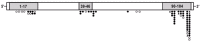
The RYR1 pathogenic variants associated with CCD identified so far are clustered in three relatively restricted regions ("hot spots"), which encode domain 1 (exons 1-17), domain 2 (exons 39-46), and domain 3 (exons 90-104) of the ryanodine receptor 1 [Treves et al 2005] (Figure 2).

Figure 2.

Figure 2.

RYR1 pathogenic variant map for CCD The three shaded mutational hot spot areas:

Although most pathogenic variants associated with CCD are clustered in the C-terminal domain 3, which comprises the transmembrane/luminal and pore-forming region of the channel, studies have shown that pathogenic variants in CCD are likewise found in domains 1 and 2, in which pathogenic variants are more commonly associated with malignant hyperthermia (see Allelic Disorders).

The most common pathogenic variants are shown in Table 3 (pdf).

Table 4 (pdf) shows the most common RYR1 pathogenic amino acid variants associated with autosomal dominant central core disease.

Normal gene product. RYR1 encodes the ryanodine receptor 1 protein, a skeletal muscle calcium-release channel located in the sarcoplasmic reticulum (SR). The functional channel is a homotetramer of 560-kd subunits; it releases calcium stored in the SR in response to membrane depolarization transduced by the dihydropyridine receptor (DHPR). The cytoplasmic domain of ryanodine receptor 1, also called the foot structure, comprises the first 4,000 amino acids that bridge the gap between the SR and the transverse tubular system. The last 1,000 amino acids from the transmembrane domain contain the pore of the channel [Tilgen et al 2001, Lehmann-Horn et al 2003].

Ryanodine receptors belong to the superfamily of intracellular Ca2+ release channels present on endoplasmic reticulum/sarcoplasmic reticulum (SR) membranes, having three different isoforms. Functional units are homotetramers of approximately 5,000 amino acids per subunit coded by 150-kb genes. RYR1, forming the SR calcium release channel, has a large hydrophilic NH2-terminal domain and a hydrophobic COOH-terminal domain containing several transmembrane domains as well as the channel pore. The 563-kd protein is predominantly expressed not only in skeletal muscle but also in human B-lymphocytes and immature murine dendritic cells.

Abnormal gene product. Alterations in the ryanodine receptor 1 protein lead to an abnormal, sustained increase in myoplasmic calcium concentration in skeletal muscle because of a "leaky channel" or uncoupling with its voltage sensor, which is encoded by the voltage-gated calcium channel gene DHPR [Nelson 2001, Wehner et al 2003].

In vitro studies suggest that a high basal activity of the mutated Ca2+ channel could explain the muscle weakness and muscle atrophy observed in persons with CCD in one family [Lynch et al 1999]. In vitro expression of ryanodine receptor 1 with a single pathogenic variant (p.Ile4898Thr) in the C-terminal transmembrane/luminal domain in HEK293 cells resulted in loss of channel activation and reduction in ryanodine binding, possibly by disrupting the ligand binding site located in the C terminus of the protein. Further analysis, however, showed that this pathogenic variant leads to a significant increase in the sensitivity of the channel to the activating effects of calcium.

The association of C-terminal pathogenic variants with clinically evident muscle weakness may be explained by the leaky-channel model and the excitation-contraction (EC) uncoupling model.

Some non-C-terminal pathogenic variants in ryanodine receptor 1 promote the leak of Ca2+ ions from the SR that may or may not be compensated by the activity of the sarco-endoplasmic reticulum Ca2+ ATPase (SERCA), resulting in elevation of resting cytosolic Ca2+ and depletion of SR Ca2+ stores.

C-terminal pathogenic variants, especially those in the pore region of ryanodine receptor 1, may directly affect the channel gating properties, resulting in an abolition of orthograde activation by the voltage-gated L-type Ca2+ channel or, in other words, EC uncoupling. However, no compensatory mechanism increases Ca2+ release as the SERCA pumps do in the leaky model. Nevertheless, the effect of pathogenic variants in the C-terminal region remains controversial because a number of pathogenic variants in this area were also shown to be "leaky." Interestingly, several pathogenic variants in RYR1 exon 102 were shown to lead to varying degrees of EC uncoupling, indicating that this region is a primary locus of EC uncoupling in CCD [Avila et al 2003].

References

Literature Cited

  • Agrawal PB, Greenleaf RS, Tomczak KK, Lehtokari VL, Wallgren-Pettersson C, Wallefeld W, Laing NG, Darras BT, Maciver SK, Dormitzer PR, Beggs AH. Nemaline myopathy with minicores caused by mutation of the CFL2 gene encoding the skeletal muscle actin-binding protein, cofilin-2. Am J Hum Genet. 2007;80:162–7. [PMC free article: PMC1785312] [PubMed: 17160903]
  • Avila G, O'Connell KM, Dirksen RT. The pore region of the skeletal muscle ryanodine receptor is a primary locus for excitation-contraction uncoupling in central core disease. J Gen Physiol. 2003;121:277–86. [PMC free article: PMC2217374] [PubMed: 12642598]
  • Boncompagni S, Rossi AE, Micaroni M, Hamilton SL, Dirksen RT, Franzini-Armstrong C, Protasi F. Characterization and temporal development of cores in a mouse model of malignant hyperthermia. Proc Natl Acad Sci U S A. 2009;106:21996–2001. [PMC free article: PMC2799858] [PubMed: 19966218]
  • Davis MR, Haan E, Jungbluth H, Sewry C, North K, Muntoni F, Kuntzer T, Lamont P, Bankier A, Tomlinson P, Sanchez A, Walsh P, Nagarajan L, Oley C, Colley A, Gedeon A, Quinlivan R, Dixon J, James D, Muller CR, Laing NG. Principal mutation hot spot for central core disease and related myopathies in the C-terminal transmembrane region of the RYR1 gene. Neuromuscul Disord. 2003;13:151–7. [PubMed: 12565913]
  • Dirksen RT, Avila G. Altered ryanodine receptor function in central core disease: leaky or uncoupled Ca(2+) release channels? Trends Cardiovasc Med. 2002;12:189–97. [PubMed: 12161072]
  • Dirksen RT, Avila G. Distinct effects on Ca2+ handling caused by malignant hyperthermia and central core disease mutations in RyR1. Biophys J. 2004;87:3193–204. [PMC free article: PMC1304789] [PubMed: 15347586]
  • Ferreiro A, Monnier N, Romero NB, Leroy JP, Bonnemann C, Haenggeli CA, Straub V, Voss WD, Nivoche Y, Jungbluth H, Lemainque A, Voit T, Lunardi J, Fardeau M, Guicheney P. A recessive form of central core disease, transiently presenting as multi-minicore disease, is associated with a homozygous mutation in the ryanodine receptor type 1 gene. Ann Neurol. 2002a;51:750–9. [PubMed: 12112081]
  • Ferreiro A, Quijano-Roy S, Pichereau C, Moghadaszadeh B, Goemans N, Bonnemann C, Jungbluth H, Straub V, Villanova M, Leroy JP, Romero NB, Martin JJ, Muntoni F, Voit T, Estournet B, Richard P, Fardeau M, Guicheney P. Mutations of the selenoprotein N gene, which is implicated in rigid spine muscular dystrophy, cause the classical phenotype of multiminicore disease: reassessing the nosology of early-onset myopathies. Am J Hum Genet. 2002b;71:739–49. [PMC free article: PMC378532] [PubMed: 12192640]
  • Fischer D, Herasse M, Ferreiro A, Barragán-Campos HM, Chiras J, Viollet L, Maugenre S, Leroy JP, Monnier N, Lunardi J, Guicheney P, Fardeau M, Romero NB. Muscle imaging in dominant core myopathies linked or unlinked to the ryanodine receptor 1 gene. Neurology. 2006;67:2217–20. [PubMed: 17190947]
  • Herasse M, Parain K, Marty I, Monnier N, Kaindl AM, Leroy JP, Richard P, Lunardi J, Romero NB, Ferreiro A. Abnormal distribution of calcium-handling proteins: a novel distinctive marker in core myopathies. J Neuropathol Exp Neurol. 2007;66:57–65. [PubMed: 17204937]
  • Ibarra M CA, Wu S, Murayama K, Minami N, Ichihara Y, Kikuchi H, Noguchi S, Hayashi YK, Ochiai R, Nishino I. Malignant hyperthermia in Japan: mutation screening of the entire ryanodine receptor type 1 gene coding region by direct sequencing. Anesthesiology. 2006;104:1146–54. [PubMed: 16732084]
  • Ilkovski B, Cooper ST, Nowak K, Ryan MM, Yang N, Schnell C, Durling HJ, Roddick LG, Wilkinson I, Kornberg AJ, Collins KJ, Wallace G, Gunning P, Hardeman EC, Laing NG, North KN. Nemaline myopathy caused by mutations in the muscle alpha-skeletal-actin gene. Am J Hum Genet. 2001;68:1333–43. [PMC free article: PMC1226120] [PubMed: 11333380]
  • Jungbluth H, Davis MR, Muller C, Counsell S, Allsop J, Chattopadhyay A, Messina S, Mercuri E, Laing NG, Sewry CA, Bydder G, Muntoni F. Magnetic resonance imaging of muscle in congenital myopathies associated with RYR1 mutations. Neuromuscul Disord. 2004;14:785–90. [PubMed: 15564033]
  • Jungbluth H, Muller CR, Halliger-Keller B, Brockington M, Brown SC, Feng L, Chattopadhyay A, Mercuri E, Manzur AY, Ferreiro A, Laing NG, Davis MR, Roper HP, Dubowitz V, Bydder G, Sewry CA, Muntoni F. Autosomal recessive inheritance of RYR1 mutations in a congenital myopathy with cores. Neurology. 2002;59:284–7. [PubMed: 12136074]
  • Jungbluth H, Zhou H, Sewry CA, Robb S, Treves S, Bitoun M, Guicheney P, Buj-Bello A, Bönnenman C, Muntoni F. Centronuclear myopathy due to a dominant mutation in the skeletal muscle ryanodine receptor (RYR1) gene. Neuromusc Disord. 2007;17:338–45. [PubMed: 17376685]
  • Kaindl AM, Ruschendorf F, Krause S, Goebel HH, Koehler K, Becker C, Pongratz D, Muller-Hocker J, Nurnberg P, Stoltenburg-Didinger G, Lochmuller H, Huebner A. Missense mutations of ACTA1 cause dominant congenital myopathy with cores. J Med Genet. 2004;41:842–8. [PMC free article: PMC1735626] [PubMed: 15520409]
  • Kossugue PM, Paim JF, Navarro MM, Silva HC, Pavanello RC, Gurgel-Giannetti J, Zatz M, Vainzof M. Central core disease due to recessive mutations in RYR1 gene: Is it more common than described? Muscle Nerve. 2007;35:670–4. [PubMed: 17226826]
  • Lehmann-Horn F, Lerche H, Jurkatt-Rott K. Skeletal muscle channelopathies: myotonias, periodic paralyses and malignant hyperthermia. In: Stalber E, ed. Clinical Neurophysiology of Disorders of Muscle and Neuromuscular Junction, Including Fatigue (Handbook of Clinical Neurophysiology), vol 2. Amsterdam, Netherlands: Elsevier; 2003:457-83.
  • Lynch PJ, Tong J, Lehane M, Mallet A, Giblin L, Heffron JJ, Vaughan P, Zafra G, MacLennan DH, McCarthy TV. A mutation in the transmembrane/luminal domain of the ryanodine receptor is associated with abnormal Ca2+ release channel function and severe central core disease. Proc Natl Acad Sci U S A. 1999;96:4164–9. [PMC free article: PMC22438] [PubMed: 10097181]
  • Monnier N, Romero NB, Lerale J, Landrieu P, Nivoche Y, Fardeau M, Lunardi J. Familial and sporadic forms of central core disease are associated with mutations in the C-terminal domain of the skeletal muscle ryanodine receptor. Hum Mol Genet. 2001;10:2581–92. [PubMed: 11709545]
  • Monnier N, Romero NB, Lerale J, Nivoche Y, Qi D, MacLennan DH, Fardeau M, Lunardi J. An autosomal dominant congenital myopathy with cores and rods is associated with a neomutation in the RYR1 gene encoding the skeletal muscle ryanodine receptor. Hum Mol Genet. 2000;9:2599–608. [PubMed: 11063719]
  • Nelson TE. Heat production during anesthetic-induced malignant hyperthermia. Biosci Rep. 2001;21:169–79. [PubMed: 11725865]
  • Norwood FL, Harling C, Chinnery PF, Eagle M, Bushby K, Straub V. Prevalence of genetic muscle disease in Northern England: in-depth analysis of a muscle clinic population. Brain. 2009;132:3175–86. [PMC free article: PMC4038491] [PubMed: 19767415]
  • Phillips MS, Fujii J, Khanna VK, DeLeon S, Yokobata K, de Jong PJ, MacLennan DH. The structural organization of the human skeletal muscle ryanodine receptor (RYR1) gene. Genomics. 1996;34:24–41. [PubMed: 8661021]
  • Quinlivan RM, Muller CR, Davis M, Laing NG, Evans GA, Dwyer J, Dove J, Roberts AP, Sewry CA. Central core disease: clinical, pathological, and genetic features. Arch Dis Child. 2003;88:1051–5. [PMC free article: PMC1719384] [PubMed: 14670767]
  • Romero NB, Monnier N, Viollet L, Cortey A, Chevallay M, Leroy JP, Lunardi J, Fardeau M. Dominant and recessive central core disease associated with RYR1 mutations and fetal akinesia. Brain. 2003;126:2341–9. [PubMed: 12937085]
  • Sato I, Wu S, Ibarra MC, Hayashi YK, Fujita H, Tojo M, Oh SJ, Nonaka I, Noguchi S, Nishino I. Congenital neuromuscular disease with uniform type 1 fiber and RYR1 mutation. Neurology. 2008;70:114–22. [PubMed: 17538032]
  • Sax TW, Rosenbaum RB. Neuromuscular disorders in pregnancy. Muscle Nerve. 2006;34:559–71. [PubMed: 16969835]
  • Sewry CA, Muller C, Davis M, Dwyer JS, Dove J, Evans G, Schroder R, Furst D, Helliwell T, Laing N, Quinlivan RC. The spectrum of pathology in central core disease. Neuromuscul Disord. 2002;12:930–8. [PubMed: 12467748]
  • Shepherd S, Ellis F, Halsall J, Hopkins P, Robinson R. RYR1 mutations in UK central core disease patients: more than just the C-terminal transmembrane region of the RYR1 gene. J Med Genet. 2004;41:e33. [PMC free article: PMC1735714] [PubMed: 14985404]
  • Tilgen N, Zorzato F, Halliger-Keller B, Muntoni F, Sewry C, Palmucci LM, Schneider C, Hauser E, Lehmann-Horn F, Muller CR, Treves S. Identification of four novel mutations in the C-terminal membrane spanning domain of the ryanodine receptor 1: association with central core disease and alteration of calcium homeostasis. Hum Mol Genet. 2001;10:2879–87. [PubMed: 11741831]
  • Treves S, Anderson AA, Ducreux S, Divet A, Bleunven C, Grasso C, Paesante S, Zorzato F. Ryanodine receptor 1 mutations, dysregulation of calcium homeostasis and neuromuscular disorders. Neuromuscul Disord. 2005;15:577–87. [PubMed: 16084090]
  • Vega AV, Ramos-Mondragón R, Calderón-Rivera A, Zarain-Herzberg A, Avila G. Calcitonin gene-related peptide restores disrupted excitation-contraction coupling in myotubes expressing central core disease mutations in RyR1. J Physiol. 2011;589:4649–69. [PMC free article: PMC3213414] [PubMed: 21825032]
  • Wehner M, Rueffert H, Koenig F, Olthoff D. Calcium release from sarcoplasmic reticulum is facilitated in human myotubes derived from carriers of the ryanodine receptor type 1 mutations Ile2182Phe and Gly2375Ala. Genet Test. 2003;7:203–11. [PubMed: 14641996]
  • Wu S, Ibarra MC, Malicdan MC, Murayama K, Ichihara Y, Kikuchi H, Nonaka I, Noguchi S, Hayashi YK, Nishino I. Central core disease is due to RYR1 mutations in more than 90% of patients. Brain. 2006;129:1470–80. [PubMed: 16621918]
  • Zhou H, Brockington M, Jungbluth H, Monk D, Stanier P, Sewry CA, Moore GE, Muntoni F. Epigenetic allele silencing unveils recessive RYR1 mutations in core myopathies. Am J Hum Genet. 2006a;79:859–68. [PMC free article: PMC1698560] [PubMed: 17033962]
  • Zhou H, Yamaguchi N, Xu L, Wang Y, Sewry C, Jungbluth H, Zorzato F, Bertini E, Muntoni F, Meissner G, Treves S. Characterization of recessive RYR1 mutations in core myopathies. Hum Mol Genet. 2006b;15:2791–803. [PubMed: 16940308]
  • Zorzato F, Yamaguchi N, Xu L, Meissner G, Muller CR, Pouliquin P, Muntoni F, Sewry C, Girard T, Treves S. Clinical and functional effects of a deletion in a COOH-terminal lumenal loop of the skeletal muscle ryanodine receptor. Hum Mol Genet. 2003;12:379–88. [PubMed: 12566385]

Chapter Notes

Revision History

  • 7 November 2019 (ma) Chapter retired: histologic diagnosis without strong genetic correlation
  • 4 December 2014 (me) Comprehensive update posted live
  • 11 May 2010 (me) Comprehensive update posted live
  • 16 May 2007 (me) Review posted live
  • 8 December 2006 (in) Original submission
Copyright © 1993-2024, University of Washington, Seattle. GeneReviews is a registered trademark of the University of Washington, Seattle. All rights reserved.

GeneReviews® chapters are owned by the University of Washington. Permission is hereby granted to reproduce, distribute, and translate copies of content materials for noncommercial research purposes only, provided that (i) credit for source (http://www.genereviews.org/) and copyright (© 1993-2024 University of Washington) are included with each copy; (ii) a link to the original material is provided whenever the material is published elsewhere on the Web; and (iii) reproducers, distributors, and/or translators comply with the GeneReviews® Copyright Notice and Usage Disclaimer. No further modifications are allowed. For clarity, excerpts of GeneReviews chapters for use in lab reports and clinic notes are a permitted use.

For more information, see the GeneReviews® Copyright Notice and Usage Disclaimer.

For questions regarding permissions or whether a specified use is allowed, contact: ude.wu@tssamda.

Bookshelf ID: NBK1391PMID: 20301565

Views

Tests in GTR by Gene

Similar articles in PubMed

See reviews...See all...

Recent Activity

Your browsing activity is empty.

Activity recording is turned off.

Turn recording back on

See more...